Dorothy Paul, DDS

Technology

Good dentistry depends on good information.

The tools we use help us see more clearly, diagnose more accurately, and explain what's going on in a way that actually makes sense to you.

Operatory at Dr. Dorothy Paul's office
Digital x-ray sensor and viewer

Diagnostic imaging

Digital x-rays

Within seconds, an enlarged image of your teeth and jaw is on the monitor. Digital x-rays help identify hidden decay, cysts, tumors, tartar buildup, and bone loss from gum disease, with far less radiation than traditional film.

Handheld radiography

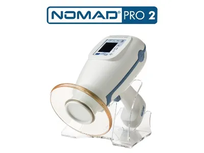
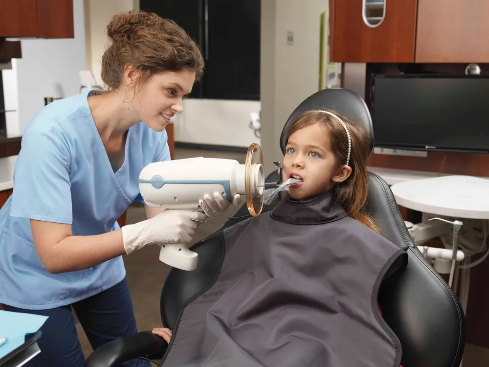
NOMAD Pro 2 handheld x-ray

Faster, more efficient intraoral x-rays. NOMAD reduces retakes by minimizing patient movement and lets the operator stay with the patient during exposure. Consistent image quality without repositioning.

NOMAD Pro 2 handheld x-ray device
Diode soft-tissue laser

Soft tissue

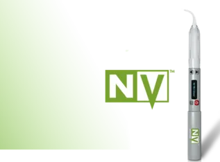
Diode soft-tissue laser

Used for frenectomies, gummy smiles, and other soft-tissue work. The laser offers more comfort during the procedure and faster healing after, compared with a traditional scalpel.

Documentation

Intraoral camera

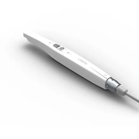
A very small camera photographs the inside of your mouth so you can see what we see: the first signs of cavities, chipped or cracked teeth, gum disease, or receding gums. Photos help you and Dr. Paul decide together.

Intraoral camera

Oral cancer screening

VELscope® oral cancer screening

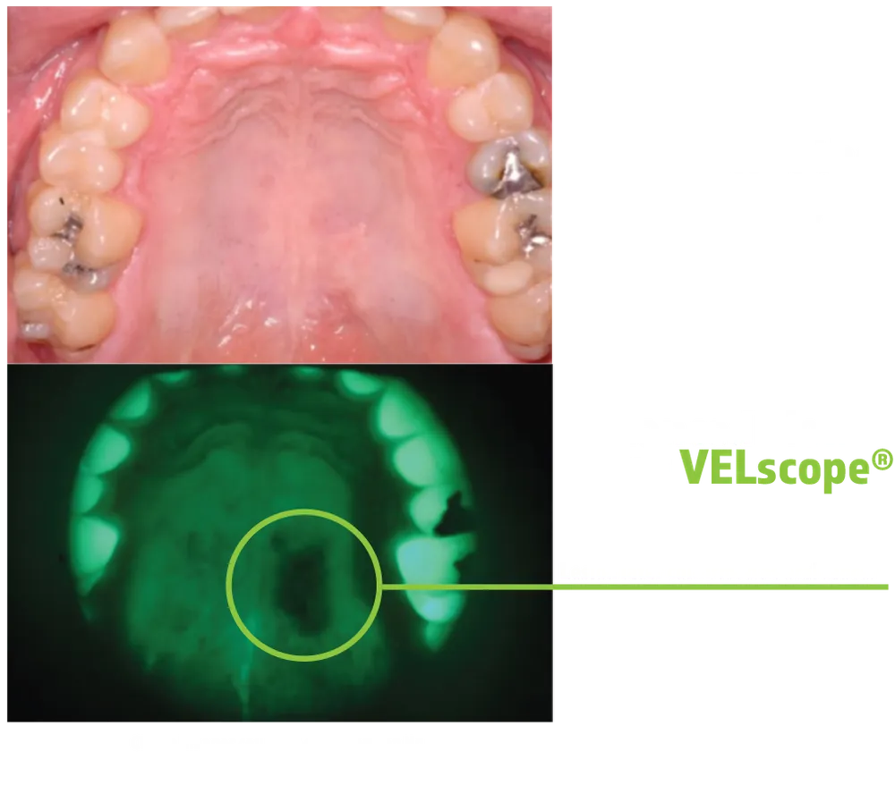
A painless, noninvasive exam that literally takes two or three minutes. The VELscope® projects blue light inside the mouth so lesions and other indicators of oral cancer, which appear darker under the specialized light, are easier to notice early. Included in every new-patient visit.

VELscope fluorescence imaging showing a lesion not visible under normal light
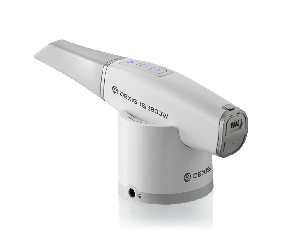
DEXIS IS 3800W intraoral scanner in its charging base

3D intraoral scanning

DEXIS IS 3800W 3D scanner

Replaces traditional putty impressions for crowns, dentures, and veneers. The scan goes directly to our Houston lab, where a more precise, fine-tuned restoration is built. Faster in the chair, more accurate on the bench.

Why the VELscope® matters

A two-minute screening that's part of every new-patient visit.

The VELscope® examination is painless and noninvasive. Dr. Paul begins with a regular visual examination of the whole lower face: glands, tongue, cheeks, palate, and teeth. Then, with protective eyewear on and the lights dimmed, the VELscope® projects blue light into the mouth so lesions and other indicators of oral cancer, which appear darker under that light, are more easily noticeable.

If anything looks concerning, Dr. Paul will refer for a biopsy. If not, another screening is performed at your next comprehensive visit. It's a small addition to the exam that catches large problems early.
Curious what a comprehensive exam looks like?

Come in and see the tools in use. Request a first visit.

Call 713-977-1010

© 2026 Dorothy Paul, DDS